The RMMR group develops new magnetic resonance imaging methods to expand the range of questions that MRI can answer. We work to make MRI more useful in challenging populations (e.g., subjects who can't stay still), expanding fast, accurate neuroimaging to the very young, old, and ill. We also work to make MRI more sensitive to lifespan changes and disease processes, helping better understand them in the lab and in life.
News
Research
Motion-Resilient Neuroimaging
Motion is a significant issue for both clinical and research MRI, making it difficult to acquire high-quality images in many populations. To address this challenge, we develop neuroimaging methods that are resilient to motion. One of our most widely used innovations is the Volumetric Navigators (vNavs) system — low-resolution volumes, acquired rapidly during an MRI scan, registered on-the-fly to track subject motion, and combined with dynamic MRI sequences that can adapt in real-time to correct for subject motion.
This method is now used in several multi-center studies, including Adolescent Brain Cognitive Development and Human Connectome Project Aging and Development. We continue to improve and expand the technology based on feedback from our collaborators. For example, we are currently refining the system for toddlers and infants as part of the HEALthy Brain and Child Development planning phase.
The image below shows the effect of motion-corrected adaptive scanning, starting with a motion-damaged scan — move the slider right to see the results of intelligently reacquiring data in periods where the subject was still, replacing motion-damaged measurements with motion-corrected ones.
Project funding: NIH (NIDA) U01DA055365 (Penn-PI: Tisdall), NIH (NIMH) R44MH121276 (Penn-PI: Tisdall), NIH (NIMH) R44MH124567 (Penn-PI: Tisdall)
Key publications:
Tisdall MD, Hess AT, Reuter M, Meintjes EM, Fischl B, van der Kouwe AJW. Volumetric navigators for prospective motion correction and selective reacquisition in neuroanatomical MRI. Magnetic Resonance in Medicine. 2012;68(2):389–399
Tisdall MD, Reuter M, Qureshi A, Buckner RL, Fischl B, van der Kouwe AJW. Prospective motion correction with volumetric navigators (vNavs) reduces the bias and variance in brain morphometry induced by subject motion. NeuroImage. 2016 Feb 15;127:11–22
Zhang Y, Aganj I, van der Kouwe AJW, Tisdall MD. Accurate High-speed 3D-Registration of EPI vNavs for Head Motion Correction. Proceedings of the 25th Annual Meeting of ISMRM; Honolulu, Hawaii. 2017. p. 3944
MRI Biomarkers of Neurodegeneration
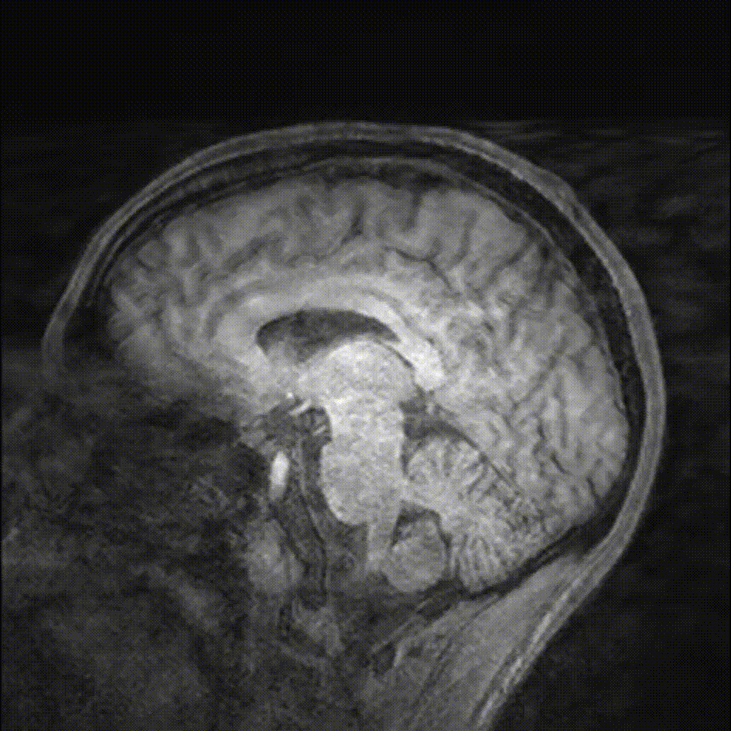
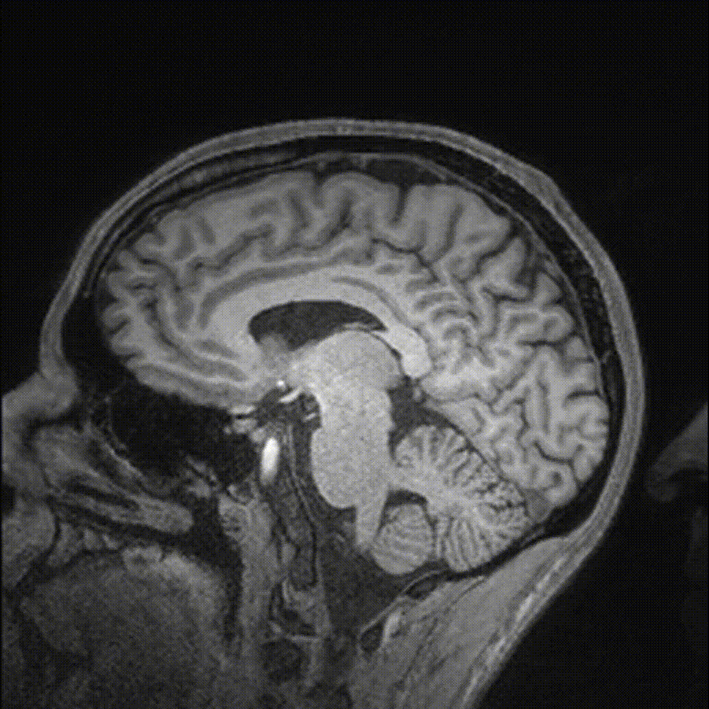
We are developing novel MRI methods, both in vivo and ex vivo to study neurodegenerative diseases. 
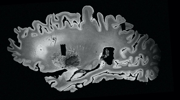
Ex vivo MRI offers a bridge between in vivo imaging and histopathology. While histopathology allows us to observe the microscopic changes in tissue structure or cellular composition caused by disease, the spatial scale of these mthods is quite limited. 
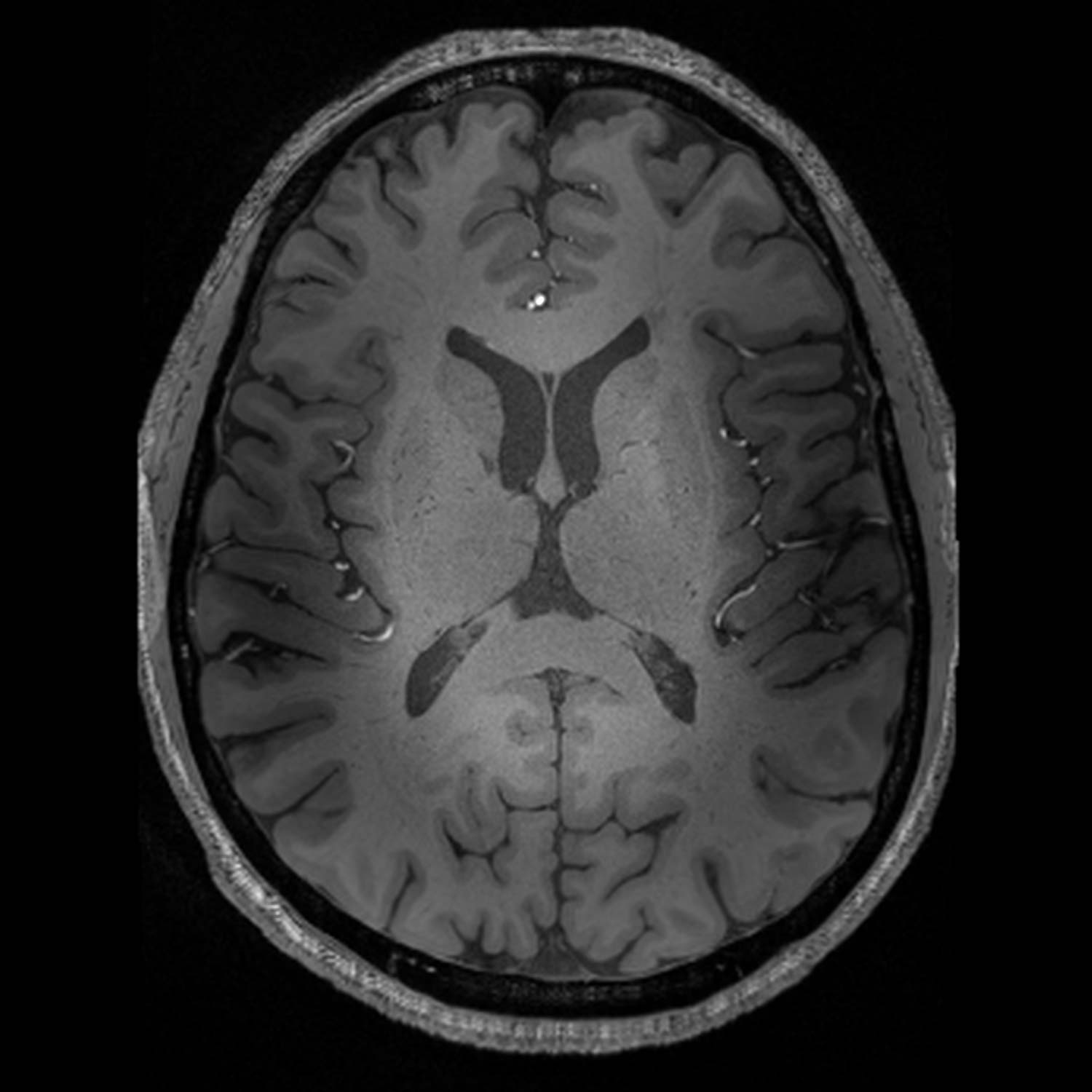
Ex vivo MRI offers an opportunity to search out mesocopic changes in tissue at the scale of whole hemispheres or even whole brains. We work closely with the 
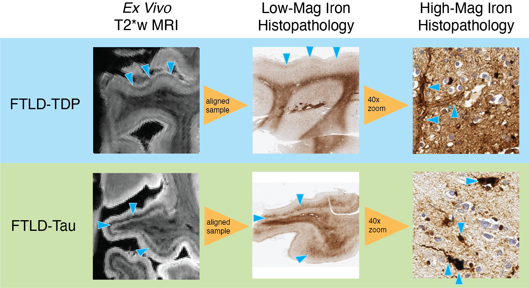
Penn Frontotemporal Dementia Center and the Penn Alzheimer's Disease Core Center to develop and evaluate ex vivo pulse sequences, studying how to maximize sensitivity to the microscopic pathological features of interest. A further translational goal of this work is to take the lessons learned from senitizing ex vivo MRI, and work to transition these methods to in vivo use as disease biomarkers. We are currently developing pulse sequences and analysis pipelines to quantify both the spatial extent of iron across the cortex, and the specific cortical laminar distribution of iron in focal disease regions.
The figure below shows the relationship between ex vivo MRI, iron stain of matched histopathology slices, and then zoomed images of the underlying iron-rich disease pathology. Note how the two rows, representing two different proteinopathies, have different pathology at the microscale, but also different distributions of iron within the cortex (black "smudges" on MRI), that could represent "imaging signatures" of each disease.
Project funding: NIH (NIA) R01AG080734 (PI: Tisdall), NIH (NIA) P01AG066597 (PIs: McMillan and Irwin)
Key publications:
MS Yao, A Van, J Gee, M Grossman, DJ Irwin, and MD Tisdall. Evaluating Echo Planar Spectroscopic Imaging with a Columnar Excitation for "Virtual Biopsies". Proceedings of the 2023 ISMRM Annual Meeting. 2023
MD Tisdall, DT Ohm, R Lobrovich, SR Das, G Mizsei, K Prabhakaran, R Ittyerah, S Lim, CT McMillan, DA Wolk, J Gee, JQ Trojanowski, EB Lee, JA Detre, P Yushkevich, M Grossman, and DJ Irwin. Ex vivo MRI and histopathology detect novel iron-rich cortical inflammation in frontotemporal lobar degeneration with tau versus TDP-43 pathology. NeuroImage: Clinical. 2022 33:102913
MD Tisdall, DT Ohm, R Lobrovich, SR Das, G Mizsei, K Prabhakaran, R Ittyerah, S Lim, CT McMillan, DA Wolk, J Gee, JQ Trojanowski, EB Lee, JA Detre, P Yushkevich, M Grossman, and DJ Irwin. T2*-weighted ex vivo whole-hemisphere 7 T MRI localizes novel focal iron-rich pathology in frontotemporal lobar degeneration. Proceedings of the 2021 ISMRM Annual Meeting. 2021. p 1920
People
Dylan Tisdall
Assistant Professor (Research), Radiology
Dylan received his PhD in computer science from Simon Fraser University, and then did postdoctoral work in MRI physics at the A. A. Martinos Center for Biomedical Imaging. He is Co-Director of the Center for Advanced MR Imaging and Spectroscopy at UPenn, and is involved in much of the activity at RMMR, but personally spends his time at the scanner developing novel methods for motion correction and ex vivo imaging.
Shraddha Pandey
Postdoctoral Fellow, Radiology
Shraddha received her PhD in Electrical Engineering from University of South Florida. She is trained in developing algorithms to accelerate the reconstruction of MRI from under sampled k-space data. She has also used deep neural network methods for MRI synthesis and analysis. At Penn she works with the Song Lab to develop and optimize MRI protocols supporting cerebrovascular imaging, including vessel wall imaging.
Karthik Prabhakaran
Staff Scientist, Neurology
Karthik received his MS in Biomedical Engineering from Wayne State University where he started his research career in MRI working on brain tumor analysis and blood oxygen saturation measurement using SWI. He came to UPenn after a stint at Princeton University where he ran the day-to-day operations of the only MRI scanner at the university and provided technical expertise for neuroimaging research studies. His current work involves the development, validation and implementation of advanced MRI protocols for neuroscience research.
Michael Yao
Bioengineering MD/PhD Candidate
Michael received his B.S. in applied physics from Caltech where he worked on engineering genetically encodable contrast agents for cancer MRI imaging. His research focuses on developing both machine learning methodology and their clinical applications for robust MRI image acquisition, reconstruction, and interpretation.
Ludwig Zhao
Bioengineering PhD Candidate
Ludwig received his B.S. in electrical engineering from UCLA and MPhil from Columbia University. He is a member of the Gottfried Laboratory and is co-advised by Dr. John Detre. His research focuses on developing methods for structural and functional imaging of the human olfactory bulb and tracts.
Alumni
Diana Zhang
PhD Candidate, Harvard University
Diana received her MEng from Harvard University, working with Dylan to develop novel motion-tracking algorithms for volumetric navigators. She is currently pursuing a PhD at Harvard University.
Marianna Gabrielyan
Scientist, Exponent
Marianna received her PhD in Physics from FIU in 2012, specializing in particle and nuclear physics. She joined Detre Lab at Penn as a postdoctoral researcher in 2017, where she performed development and validation of perfusion phantom for arterial spin labeled (ASL) MR imaging. She also worked with RMMR on pulse sequence implementation for gradient system calibration of MR scanners using Gradient Impulse Response Functions (GIRFs). In 2021 she joined Exponent as a Scientist.
Alan Chu
Neuroradiologist
Alan received his MD and PhD in Biomedical Engineering from the University of Michigan, and completed his residency and Neuroradiology fellowship at the UPenn. During his residency and fellowhip, Alan received an RSNA award to research dynamic shim correction and detection of iron within the brain, particularly in the context of neurodegeneration. In 2022 he became a practicing neuroradiologist.
Join the Group

We have several open postdoctoral positions for candidates with backgrounds in bioengineering, computer science, physics, and related fields. Check out our ads for positions in both motion-resilient neuroimaging and
biomarker development in frontotemporal dementia.

We are open to graduate students interested in rotations with the group. Please contact Dylan Tisdall (mtisdall@pennmedicine.upenn.edu) to discuss available projects.